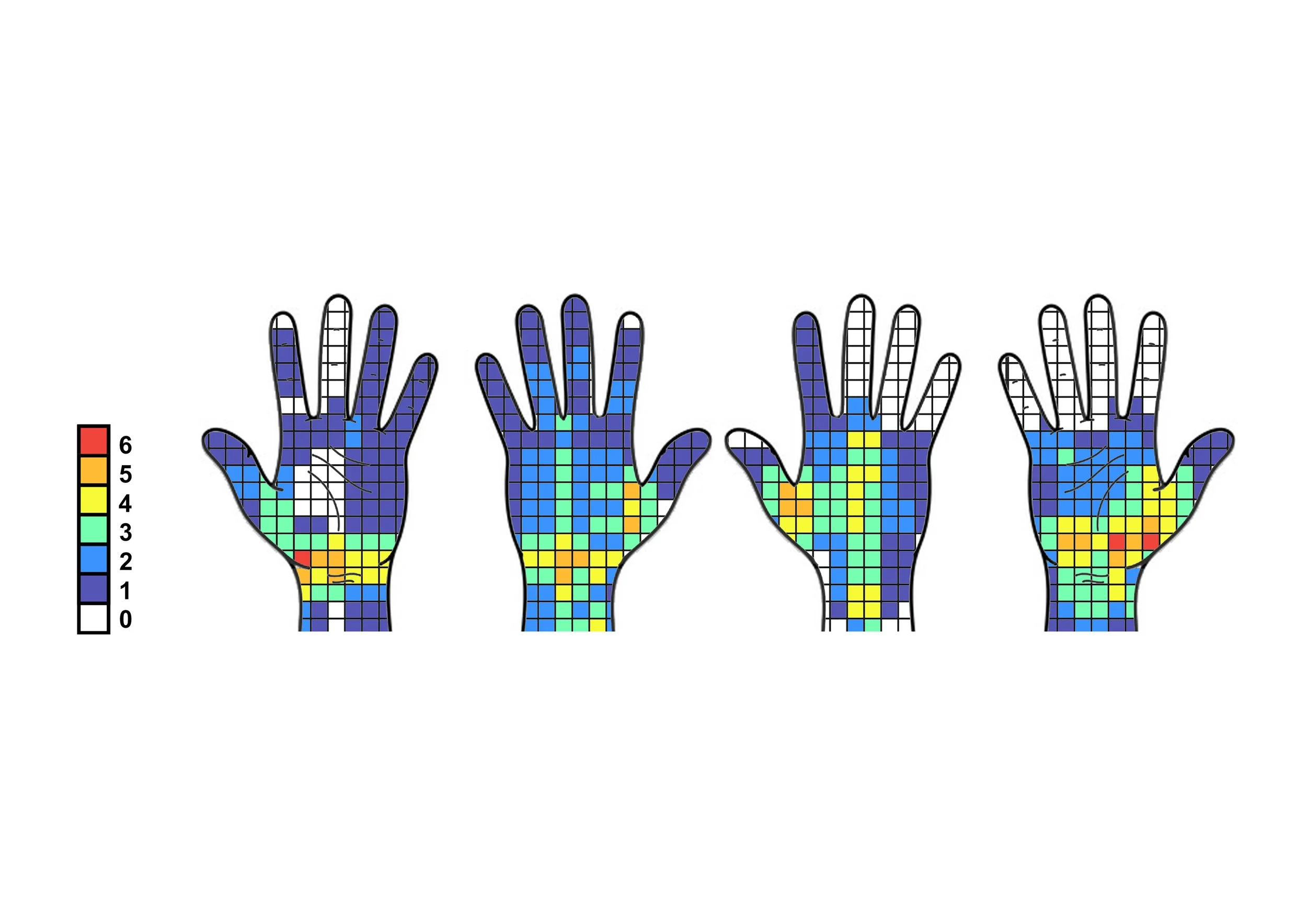
Our research has indicated that the number of imaging procedures carried out by a photographer over the duration of their career, may be the key to calculating the risk factors for developing Musculoskeletal Disorders.
Advances in camera technology have made imaging the retina an invaluable part of the patient’s visit, and because of this, a high proportion require a scan and a photograph, as part of their appointment. In 2004, the imaging department at Oxford Eye Hospital photographed between 10–20 patients a day, documenting the back and front of the eye using Colour Fundus Photography (CFP), Fluorescein Angiography (FA), Indocyanine Green Angiography (ICGA), Fundus Auto-Fluorescence (FAF), and Anterior Segment Photography. It was the introduction of Optical Coherence Tomography (OCT), a non-invasive test that increased the requests for imaging dramatically. The number of patients in Oxford that currently required imaging on an average day, range between 150–200. These patients are imaged as walk round requests from the clinic, with other procedures (FA, ICGA), booked into allocated time slots so the appropriate number of patients can be photographed in line with the staffing levels. This is calculated by estimating that the average time to perform a scan and photograph is 10 minutes, allowing the photographer 5 minutes for each procedure. Since the introduction of OCT the reliance on imaging has created a repetitive cycle, where during an average day, the photographer is constantly scanning and photographing the retina. I noticed that pain and discomfort was reported by a percentage of the imaging team in Oxford while performing these procedures. The question of whether this is related to the repetitive actions, awkward positions inherent with performing the tests, and high procedure output was asked, and whether the long-term use of the equipment could lead to developing Musculoskeletal (MSK) problems. If you would like to find out more about the results please read my paper titled An exploratory ergonomic evaluation of musculoskeletal risks for ophthalmic photographers who use ophthalmic imaging equipment plus user equipment trials. In 2023 the paper was awarded the Taylor and Francis Reader Choice Award.
MSK presentation
The results from the paper were presented at the Institute of Medical Illustrators conference in Glasgow. After the talk I was approached by several photographers who thanked me for highlighting problems they were encountering and used this evidence to feedback to their employers.