Maps are incredibly useful for navigation, planning, and displaying complex data.
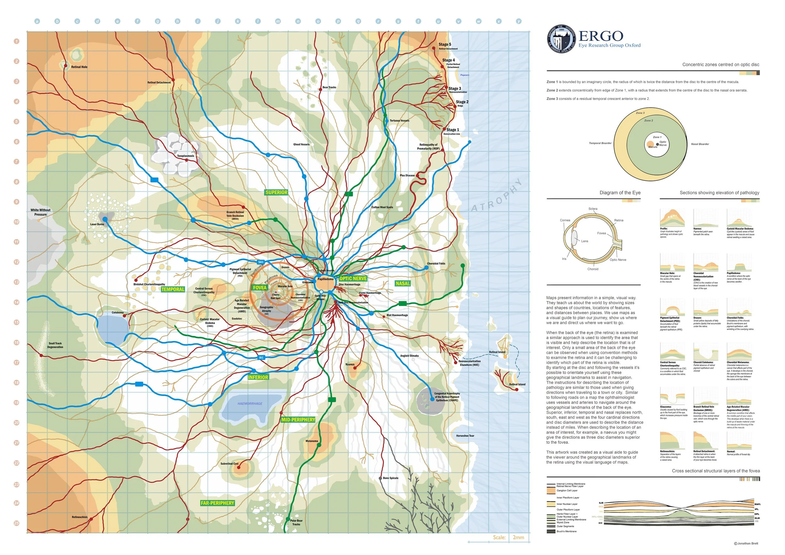
Maps present information in a simple, visual way. They teach us about the world by showing sizes and shapes of countries, locations of features, and distances between places. We use maps as a visual guide to plan our journey, show us where we are and direct us where we want to go.
When the back of the eye (the retina) is examined a similar approach is used to identify the area that is visible and help describe the location that is of interest. Only a small area of the retina can be observed when using conventional methods to examine the retina and it can be challenging to identify which part of the retina is visible.
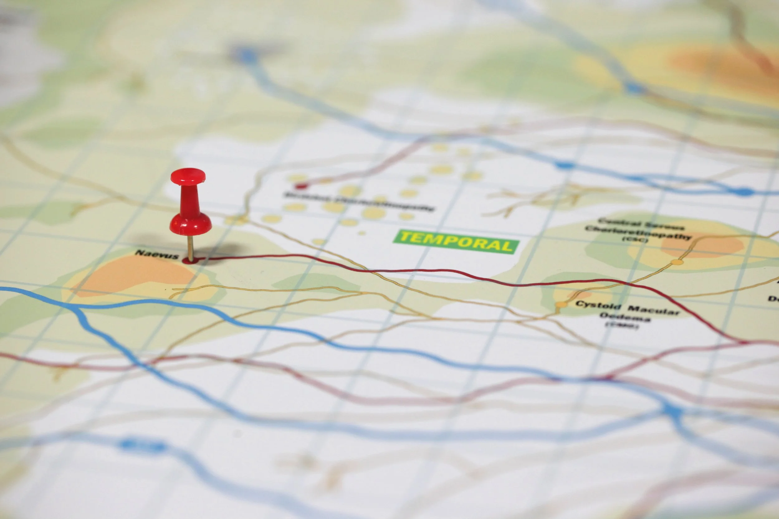
By starting at the optic nerve and following the vessels it’s possible to orientate yourself using these geographical landmarks to assist in navigation. The instructions for describing the location of pathology are similar to those used when giving directions when traveling to a town or city. Similar to following roads on a map the ophthalmologist uses vessels and arteries to navigate around the geographical landmarks of the back of the eye. Superior, inferior, temporal and nasal replaces north, south, east and west as the four cardinal directions and disc diameters are used to describe the distance instead of miles. When describing the location of an area of interest, for example, a naevus you might give the directions as three disc diameters superior to the fovea.
This artwork was created as a visual aide to guide the viewer around the geographical landmarks of the retina using the visual language of maps.
The Retinal Road map was submitted as part of the 2021 Institute of Medical Illustrators annual awards where I was fortunate to be presented with the Gabriel Donald Award.
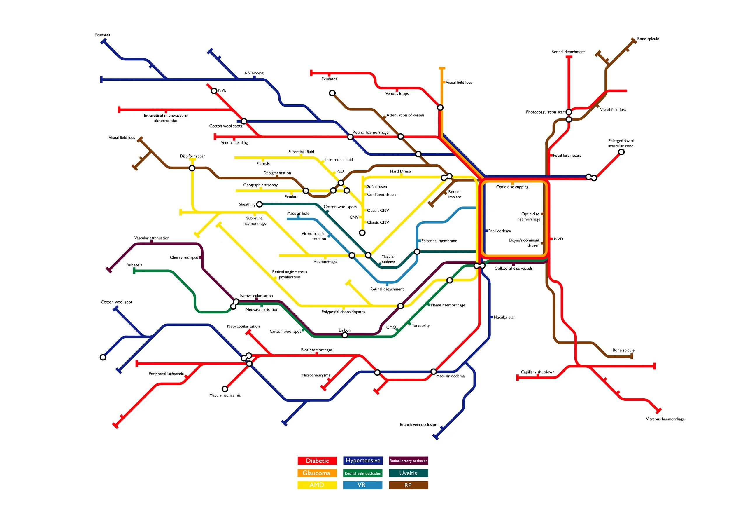
Underground Map
I have been interested in maps and how to display complex information since my training as a graphic designer. Working within healthcare has given me opportunities to explore this further. Before I created the road map I worked on a diagram of the eye using the visual language of the London Underground Map.
The underground map was designed by Harry Beck, an electrical draughtsman who created the diagram in 1931, basing it on circuit diagrams to focus on clarity and connections rather than geographical accuracy.
I replaced the lines with common eye disorders and changes the stops to pathology that may be present with each of these conditions and placed them in the geographic location they may be found.
FFA world map
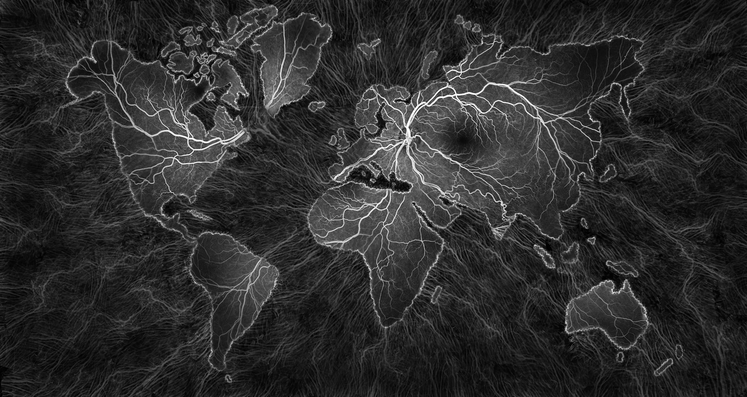
While experimenting with the FFA montage setting on the Heidelberg I noticed the edges of the remaining healthy retina looked like a coastline and the remaining healthy retina looked like an island. This led on to another map related design. This artwork was submitted to the 2019 Institute of Medical Illustrators awards where I was presented with the Gabriel Donald award.